Welcome to MITR Hospital
Kidney Cancer vs Kidney Cyst: What Is the Difference?
Finding an abnormal growth on the kidney during an ultrasound or CT scan can be worrying. Many people immediately wonder whether it is cancer. However, not every kidney growth is cancerous. A kidney cyst is often a harmless, fluid-filled sac, while kidney cancer develops when abnormal cells grow uncontrollably in kidney tissue.
Understanding the difference between a kidney cyst and kidney cancer is important because the diagnosis determines whether you need monitoring, further investigations, or treatment. Some cysts require no treatment at all, whereas suspicious kidney masses need careful evaluation by a specialist.
For people searching for Kidney Cancer treatment in Navi Mumbai, knowing how these conditions differ can help them understand their scan reports and make informed decisions about the next steps.
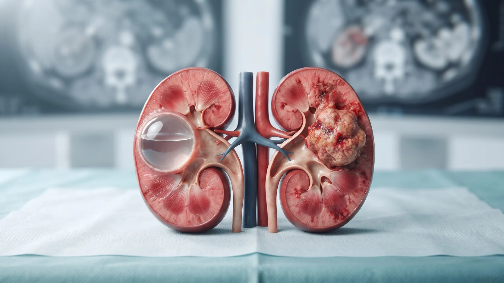
What Is a Kidney Cyst?
A kidney cyst is a round or oval sac filled with fluid that develops in or on the kidney. Simple kidney cysts become more common with age and are frequently discovered incidentally during imaging tests performed for unrelated reasons.
Most simple kidney cysts are benign, meaning they are not cancerous. They generally do not affect kidney function and may not cause any symptoms.
Kidney cysts can be broadly described as follows:
1. Simple Kidney Cysts
Simple cysts typically have thin, smooth walls and contain fluid. They have a characteristic appearance on imaging and are usually harmless.
In most cases, simple kidney cysts do not require treatment. Your doctor may recommend monitoring if there is uncertainty about the diagnosis or if the cyst causes symptoms.
2. Complex Kidney Cysts
Complex cysts have features that differ from those of simple cysts. They may contain internal divisions, thicker walls, calcifications, or solid components.
These features do not automatically mean that a cyst is cancerous. However, complex cysts may require further imaging and follow-up to assess their risk.
Doctors may use the Bosniak classification system to categorise certain cystic kidney lesions according to their appearance on contrast-enhanced CT or MRI scans. The category helps guide decisions about monitoring or treatment.
What Is Kidney Cancer?
Kidney cancer occurs when abnormal cells grow uncontrollably within kidney tissue. The most common type in adults is renal cell carcinoma.
Unlike a typical simple kidney cyst, a kidney cancer may appear as a solid mass. Some kidney cancers can also have cystic features, which is why imaging findings must be interpreted carefully.
Kidney cancer may be discovered during a scan performed for another reason. In its early stages, it may not cause noticeable symptoms.
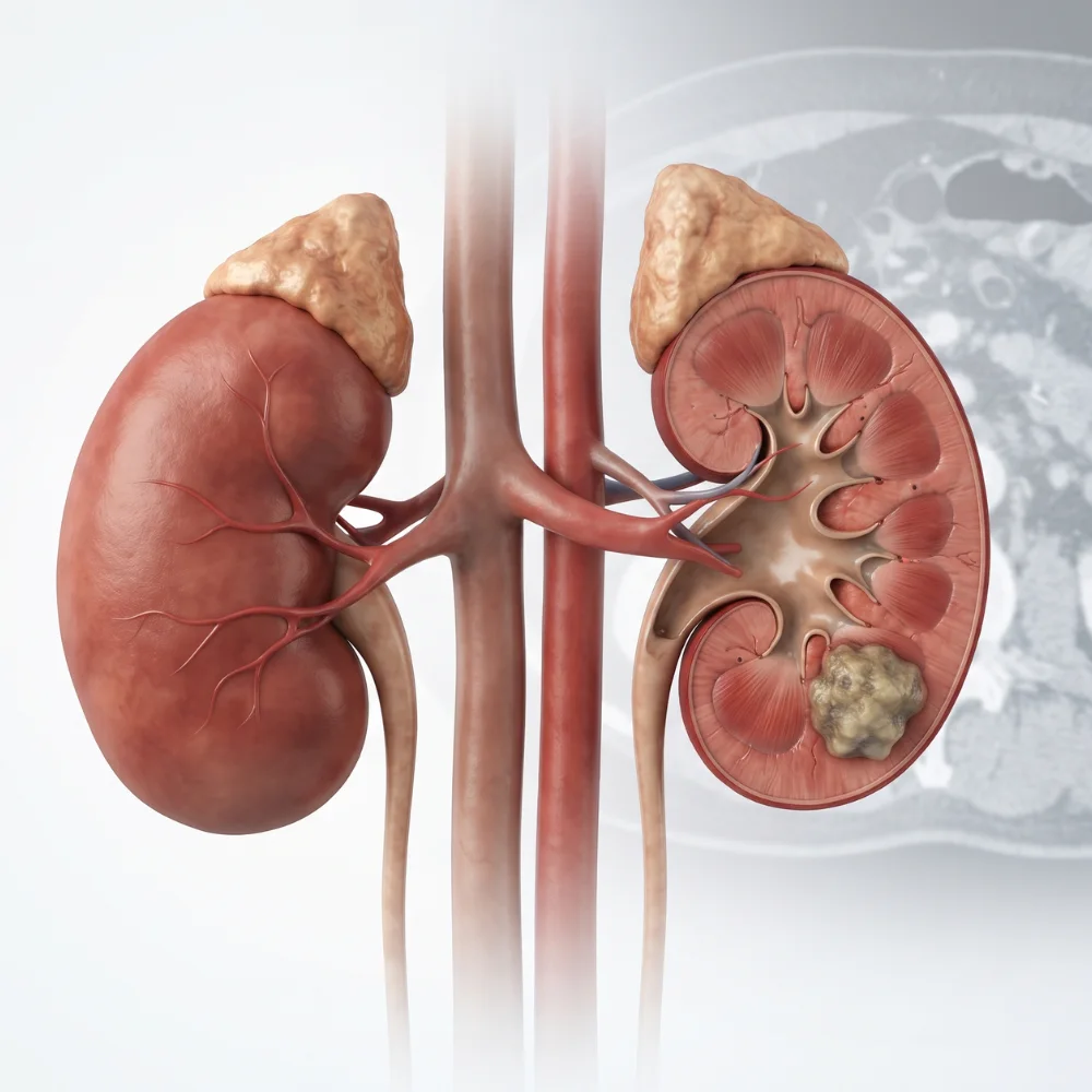
As the condition progresses, some people may develop blood in the urine, persistent pain in the side or back, an abdominal lump, unexplained weight loss, or ongoing fatigue. These symptoms can also occur with other medical conditions and do not confirm cancer on their own.
Early assessment of a suspicious kidney mass helps doctors determine the appropriate management plan.
Kidney Cancer vs Kidney Cyst: Key Differences
The main difference is that a simple kidney cyst is generally a benign fluid-filled sac, whereas kidney cancer involves cancerous cells. However, some complex cysts can have a risk of malignancy, and imaging is needed to distinguish between them.
| Feature | Simple kidney cyst | Kidney cancer |
|---|---|---|
| Nature | Usually benign and fluid-filled | Cancerous growth of kidney cells |
| Appearance | Usually thin-walled with clear fluid | Often a solid mass; some lesions have cystic features |
| Symptoms | Usually no symptoms | Often silent early on; symptoms may develop later |
| Effect on kidney function | Usually does not affect function | Depends on size, location, extent and treatment |
| Diagnosis | Usually identified through imaging | Imaging, clinical evaluation and sometimes biopsy |
| Treatment | Often no treatment is needed | May require surgery, active surveillance or other cancer treatment |
| Follow-up | Depends on the cyst's appearance and symptoms | Based on diagnosis, stage and treatment plan |
Important: Symptoms alone cannot reliably distinguish a kidney cyst from kidney cancer. The appearance of the lesion on appropriate imaging is often central to the assessment.
What Symptoms Should You Look Out For?
Symptoms of a Kidney Cyst
Most simple kidney cysts do not cause symptoms. When a cyst becomes large or develops a complication, possible symptoms include:
- Dull pain in the side, back, or upper abdomen
- A feeling of pressure or discomfort
- Fever and tenderness if the cyst becomes infected
- Blood in the urine in certain cases
- Problems with urine drainage if a cyst presses on nearby structures
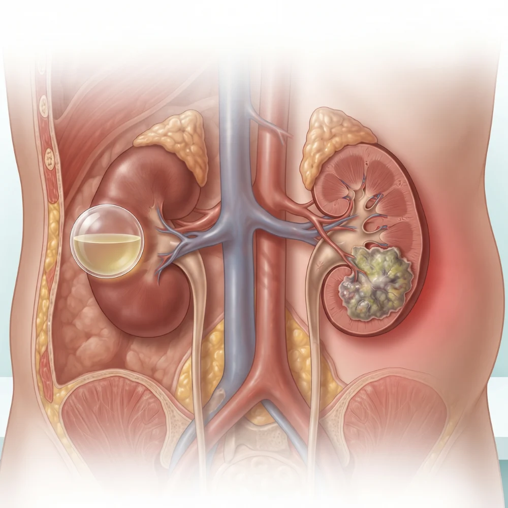
These symptoms do not necessarily indicate cancer, but they should be evaluated by a healthcare professional.
Symptoms of Kidney Cancer
Kidney cancer may not cause symptoms in its early stages. When symptoms occur, they can include:
- Blood in the urine, which may appear pink, red, or brown
- Persistent pain in the side or back
- An unusual lump or swelling in the abdomen
- Unexplained weight loss
- Persistent fatigue
- Ongoing fever without an obvious cause
Many of these symptoms have other possible explanations. However, visible blood in the urine, persistent unexplained pain, or an abdominal lump should not be ignored.
Seek urgent medical care if you experience severe pain, heavy bleeding, or symptoms accompanied by significant weakness or feeling seriously unwell.
How Do Doctors Diagnose a Kidney Cyst or Kidney Cancer?
The diagnostic process depends on the scan findings, your symptoms, medical history, and other risk factors. The goal is to identify the nature of the kidney lesion and determine whether further investigation is needed.
1. Ultrasound
An ultrasound can help identify a fluid-filled cyst and distinguish a typical simple cyst from a lesion that needs further evaluation.
However, ultrasound may not provide enough detail to characterise every kidney mass. Your doctor may recommend additional imaging if the findings are unclear.
2. CT Scan
A CT scan provides detailed images of the kidneys and surrounding structures. When appropriate, a contrast-enhanced CT can help assess whether a lesion contains solid tissue that takes up contrast, along with other features relevant to diagnosis.
The findings help doctors evaluate suspicious masses and plan treatment when necessary.
3. MRI Scan
An MRI may be recommended when a lesion needs further characterisation or when a CT scan is unsuitable or inconclusive.
Your doctor will select the imaging test based on the lesion's features, your kidney function, and other clinical considerations.
4. Blood and Urine Tests
Blood tests can assess kidney function and general health. Urine tests may help identify blood or other abnormalities.
These tests cannot independently confirm or exclude kidney cancer, but they provide useful information as part of the overall assessment.
5. Kidney Biopsy
A biopsy involves collecting a small tissue sample for examination. It may be recommended in selected cases when the result could influence management.
A biopsy is not necessary for every kidney mass. Your specialist will determine whether it is appropriate based on the imaging findings and treatment options.
What Is the Treatment for a Kidney Cyst?
Treatment depends on whether the cyst is simple or complex, whether it causes symptoms, and whether it affects kidney function.
Monitoring
A typical simple cyst that causes no symptoms may not require treatment. Depending on the findings, your doctor may recommend no further testing or periodic imaging.
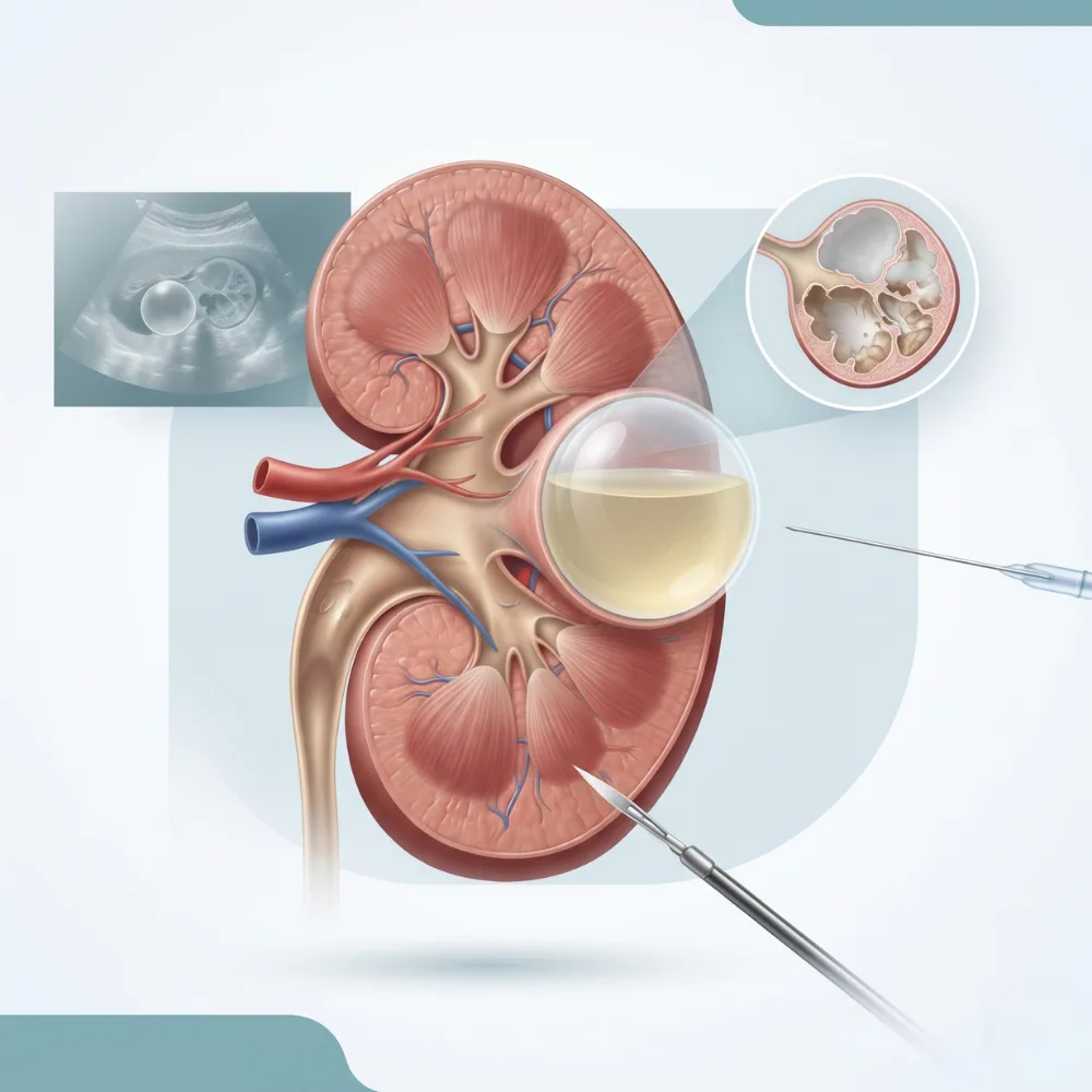
Cyst Drainage or Sclerotherapy
If a cyst causes symptoms, a doctor may consider draining it and using a substance to reduce the likelihood of fluid returning. This approach is suitable only for selected cysts.
Laparoscopic Surgery
A large or persistently symptomatic cyst may sometimes require a minimally invasive procedure to address the cyst wall or relieve pressure.
Complex cysts that have concerning imaging features need a different assessment. Their management may involve active surveillance, additional investigations, or treatment depending on their risk category.
What Are the Treatment Options for Kidney Cancer?
Treatment depends on the size and location of the tumour, whether it has spread, kidney function, overall health, and individual preferences.
People looking for Kidney Cancer treatment in Navi Mumbai should seek an assessment from a urologist experienced in evaluating kidney masses and discussing appropriate treatment options.
1. Active Surveillance
Some small kidney masses may be monitored with scheduled imaging rather than treated immediately. This approach may be appropriate for selected patients after careful assessment.
The decision depends on the lesion's characteristics, the person's overall health, and the potential benefits and risks of intervention.
2. Partial Nephrectomy
A partial nephrectomy removes the tumour and a margin of surrounding tissue while preserving the remaining healthy kidney.
When technically and medically appropriate, this approach can help preserve kidney function while treating the cancer.
3. Radical Nephrectomy
A radical nephrectomy involves removing the affected kidney and surrounding tissues as required by the tumour's location and extent.
It may be recommended when the tumour cannot be safely or effectively removed with a partial nephrectomy.
4. Ablation Procedures
Selected small kidney tumours may be treated using techniques such as cryoablation or thermal ablation. These methods destroy targeted tissue and may be considered for certain patients who are not suitable candidates for surgery or when other clinical factors favour them.
5. Systemic Treatment
For kidney cancer that has spread or requires treatment beyond local surgery, options may include immunotherapy or targeted medicines. The appropriate treatment depends on the cancer type, stage, and the patient's overall condition.
Not every patient needs all these treatments. The treating team develops a plan based on the individual diagnosis.
Can A Kidney Cyst Turn Into Cancer?
A typical simple kidney cyst is not considered a cancerous growth and generally does not turn into kidney cancer.
However, some complex cystic lesions have features associated with an increased risk of malignancy. This is why it is important to understand the exact wording of an imaging report rather than relying only on the term "kidney cyst."
If your report mentions a complex cyst, septations, a thickened wall, a solid component, or a Bosniak category, ask your doctor what the finding means and whether additional imaging or follow-up is required.
Do not assume that every complex cyst is cancerous. Equally, do not ignore a lesion simply because the report uses the word "cyst."
Who Is at Risk of Kidney Cancer?
Several factors are associated with an increased risk of kidney cancer, although having one or more risk factors does not mean that someone will develop the disease.
These factors include:
- Smoking
- Excess body weight
- High blood pressure
- Certain inherited conditions and a family history of kidney cancer
- Some long-term medical or occupational exposures
Kidney cysts are common, especially with increasing age, and most simple cysts are unrelated to kidney cancer.
If you have a family history of kidney cancer, a known inherited cancer syndrome, or a suspicious kidney mass, discuss the appropriate assessment and follow-up with your doctor.
When Should You Consult a Urologist?
Arrange a medical evaluation if:
- An ultrasound or CT scan identifies a complex cyst or an unexplained kidney mass.
- You notice blood in your urine, even if it happens only once.
- You experience persistent pain in your side or back without a clear explanation.
- Your imaging report recommends further investigation or follow-up.
- You have a known kidney lesion and are unsure whether monitoring is still required.

If your scan has already identified a kidney lesion, bring the complete report and previous images to your appointment when possible. Comparing earlier and current scans can help your doctor assess whether the lesion has changed.
Kidney Cancer Treatment in Navi Mumbai: Why Specialist Evaluation Matters
A kidney cyst and kidney cancer can require very different approaches. While many simple cysts need no treatment, a suspicious kidney mass may require further imaging, monitoring, a biopsy in selected cases, or surgery.
If you are looking for Kidney Cancer treatment in Navi Mumbai, MITR Urology Associates can be a point of contact to discuss kidney-related concerns and understand the next steps in your evaluation. The appropriate care plan should be based on your scan findings, kidney function, overall health, and specialist assessment.
Early evaluation does not mean that a suspicious finding is definitely cancer. It helps establish what the lesion is and whether treatment is necessary.
Frequently Asked Questions
No. A simple kidney cyst is usually a benign, fluid-filled sac, whereas kidney cancer involves abnormal cancerous cells. Some complex cysts can have concerning features and require further evaluation.
Ultrasound can often identify a typical simple cyst. However, some lesions need a CT scan or MRI for more detailed assessment. The appropriate test depends on the initial findings.
No. Most simple kidney cysts do not need treatment if they cause no symptoms or complications. Treatment may be considered for symptomatic cysts or lesions with concerning features.
A complex kidney cyst has imaging features beyond those of a simple fluid-filled sac. Depending on its appearance and classification, it may need further imaging, monitoring, or treatment.
Yes. Kidney cancer may be discovered during imaging performed for an unrelated reason. The absence of symptoms does not always exclude a concerning kidney lesion.
No. Treatment depends on the tumour's size, location, stage, and the patient's health. Options may include active surveillance for selected small masses, partial or radical nephrectomy, ablation, or systemic treatment.
Consult a urologist if your imaging report identifies a suspicious kidney mass, a complex cyst requiring evaluation, or if you experience symptoms such as blood in the urine or persistent unexplained side pain. A specialist can determine whether further testing or treatment is needed.
Conclusion
The difference between kidney cancer and a kidney cyst lies mainly in the nature of the lesion and its features on imaging. Most simple kidney cysts are harmless and do not require treatment, while complex cysts and suspicious kidney masses may need further investigation.
If a scan has identified an abnormality in your kidney, avoid jumping to conclusions or delaying recommended follow-up. Consult a urologist to understand the findings and discuss the most appropriate next steps.
For those seeking Kidney Cancer treatment in Navi Mumbai, MITR Urology Associates can help you discuss your kidney health concerns and the evaluation or treatment options that may be appropriate for your condition.
Medical disclaimer: This article is for educational purposes only and is not a substitute for professional medical advice, diagnosis, or treatment.
Consult a Kidney Specialist
Concerned about a kidney cyst or kidney mass on your scan? Book an appointment with the experienced urology team at MITR Urology Associates to receive comprehensive evaluation, advanced diagnostics, and personalized care.
Take Charge of Your Health Today
Book a Consultation with a SpecialistCopyright © All Rights Reserved, By MITR Urology Associates